Monographs Banner

Met-enkephalin

MENK
Met-enk
Metenkephalin
Methionine-enkephalin

Opioid Growth-Factor (OGF)

Contents

Sequence:

Tyr-Gly-Gly-Phe-Met

Molar Weight:

573.6611 g/mol

Molecular Formula:

C27H5N35SO7


Most Frequent Uses:
  • Antinociception – pain management support
  • Immune support – cancer/AIDS adjuvant; post-vaccine immune support
  • Stress response support
  • Substance use disorders
  • Metabolic Syndrome – improved white fat browning
Dosage:
  • Met-enkephaplin has a very short half-life (t1/2 = < 2 minutes)[i]
  • General Dosage:
    • 5 mg per administration given 3x/week (15mg/week).
    • Max 10 mg SubQ BID
    • IV infusion 250 mcg/kg over 30 min weekly

[i] Hartvig P, et al. Kinetics of four 11C-labelled enkephalin peptides in the brain, pituitary and plasma of rhesus monkeys. Regul Pept. 1986;16(1):1-13.

Safety and Potential Side Effects/Contraindications:
  • Met-enkephalin peptides administered subcutaneously are reported safe and efficacious in recommended dosages.
  • As with all injections, redness, and pain at the site of injection may be present.
Description

Enkephalins are endogenous pentapeptides involved in regulating pain sensation (nociception). Structurally, the enkephalins are pentapeptides that are distinguished into two subgroups by their carboxy-terminal amino acids, leucine, or methionine. Consequently, the enkephalins are either classify as met-encephalins and leu-encephalins, respectively:

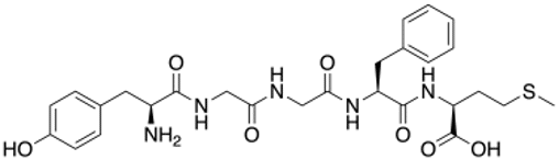
  • The met-enkephalinspresent the amino acid sequence Tyr-Gly-Gly-Phe-Met.
  • The leu-enkephalinspresent the amino acid sequence Tyr-Gly-Gly-Phe-Leu.

Enkephalin peptides act as neurotransmitters and neuromodulators throughout the nervous system and various end-organ targets.[i] Additionally, research has found that met-enkephalin has an essential role in cell proliferation and tissue organization during development. 

Early Development of Enkephalins

During the 1980s, researchers discovered that the endogenous opioid system, and met-enkephalin, in particular, has a role in regulating cell proliferation in both normal and neoplastic cell lines. Met-enkephalin, in this context, has the name OGF, and its target receptor is the OGF receptor (OGFr). Notably, OGFr is significantly different in its structure and molecular composition as compared to the classical mu, delta, and kappa opioid receptors. The principal mechanism of action is through the upregulation of p16 and p21 cyclin-dependent kinases, which serve to stall the progression of the cell replication cycle from the G0/G1 phase to the S phase. The system is tonically active, and OGFr protein and gene expression are present in most proliferating cell lines.[9] 

Organ systems involved include:

  • Nervous systems – CNS, PNS, ANS
  • Cardiac systems
  • Respiratory system
  • Gastrointestinal system
  • Endocrine system
  • Immune system

Function of Enkephalins[ii]

The expression of enkephalins and their target opioid receptors have a wide distribution throughout the central, peripheral, and autonomic nervous systems, multiple organ systems, as well as endocrine tissues and their target organs. The various effects of enkephalins are best understood by considering a small sample of these experimentally studied systems. The extensive literature on enkephalins physiological effects includes but is not limited to its role in analgesia, angiogenesis, blood pressure regulation, embryonic development, feeding, hypoxia, limbic system modulation (emotional conditions), memory processes, neuroprotection, peristalsis, pancreatic secretion, wound repair, respiratory control, and hepatoprotective mechanisms. Again, leu-enkephalin seems to be involved in the control of the gonadal function. The main functions concern analgesia, stress response regulation, and peristalsis modulation.

Functions include:

  • Analgesia
  • Stress response regulation (improves release of corticotropin-releasing factor (CRF))
  • Peristalsis – slow GI motility by alerting neuronal excitability

Mechanisms[iii]

Enkephalins exert their physiological effect through specific opioid receptors, which have a broad distribution in the body. Three major classes of opioid receptors exist and are named mu (mainly expressed in the CNS), delta (equally expressed in the SNC and spinal cord), and kappa (expressed primarily in the spinal cord). The fourth class of opioid receptors called nociceptin was discovered in 1994 but is not considered to be part of the aforementioned tripartite group, which is often referred to as the classical opioid receptors.

Enkephalins have the highest affinity for the delta-opioid receptor, followed by the mu-opioid receptor, and exhibit low affinity for the kappa-opioid receptor.

Opioid receptors are in the family of G-protein coupled receptors recognized by their seven membrane-spanning motifs with approximately 60% sequence homology. Their extracellular domains, which determine their selectivity, reported between 34 to 49% when comparing sequence similarity. The significant inhibitory effects of enkephalins are mediated by reducing K+ and Ca2+ influx. Signal transduction begins with ligand binding, which causes dissociation of the Gα and Gβγ subunits. The Gα subunit directly interacts with inward rectifying potassium channels causing cellular hyperpolarization. The Gα subunit also inhibits adenylyl cyclase activity, which decreases the formation of cAMP, thus reducing the cAMP-dependent Ca2+ influx. The Gβγ further reduces calcium influx by directly binding to various classes of Ca2+ channels.

Drug Abuse

The role of the endogenous enkephalinergic system (e.g., enkephalin peptides and receptors they bind to) in modulating the reward pathway and reward-related behaviors because (1) there is widespread synthesis and release of enkephalins in the reward pathway and (2) the receptor targets of enkephalin are also widely distributed throughout the reward circuitry, namely the mesolimbic and nigrostriatal pathways.[iv] Enkephalins can potentially be used for substance or other reward-based addictions.

Met-Enkephalin (MENK)

Methionine enkephalin (Met-enkephalin, MENK or Met-enk), was originally discovered by Hughes in 1975.[v] It is an endogenous opioid, derived from pre-enkephalin and has the amino acid sequence of Tyr-Gly-Gly-Phe-Met. MENK is found in the blood at low concentrations and is present in all parts of the nervous system. Studies into the neuroanatomic relationship of MENK have reported higher levels of MENK in the caudate nucleus, globus pallidus, putamen and substantia nigra by radioimmunoassay. As a member of the endogenous opiate family, MENK is well-known as a long-lasting analgesic, and has an important role in modulating pain sensitivity.[vi]

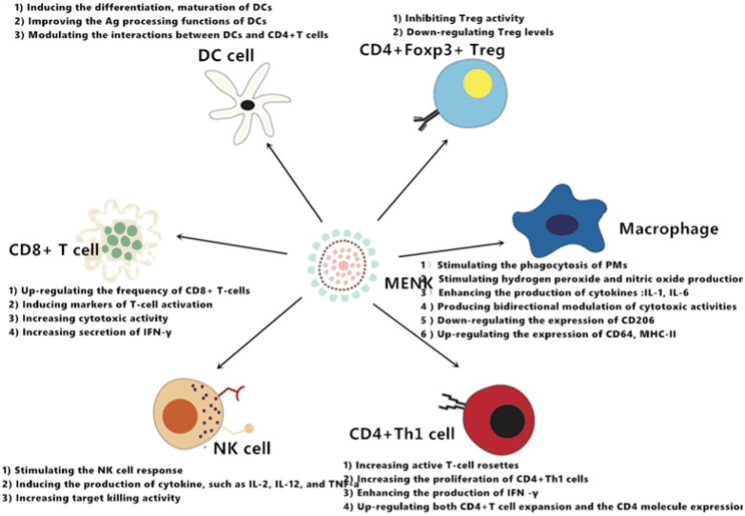
There are increasing numbers of reports supporting the observation that MENK, is involved in a regulatory loop between the neuroendocrine and immune systems and has an immune modulatory role.

Met-Enkephalin and Immunity

Met-enkephalin (MENK), an endogenous neuropeptide has a crucial role in both neuroendocrine and immune systems.[vii] MENK is believed to have an immunoregulatory activity to have cancer biotherapy activity by binding to the opioid receptors on immune and cancer cells. Clinical studies in cancer patients have reported that MENK activates immune cells directly and by inhibiting regulatory T-cells (Tregs).[viii] MENK may also change the tumor microenvironment by binding to opioid receptor on or in cancer cells. All of these mechanisms of action have biologic significance and potential for use in cancer immunotherapy. Furthermore, they reveal a relationship between the endocrine and immune systems. Due to the apparent role of MENK in cancer therapy we reviewed herein, the research undertaken with MENK in recent years; which has advanced our understanding of the role MENK has in cancer progression and its relationship to immunity, supporting MENK as a new strategy for cancer immunotherapy.

MENK has been reported to exert anti-influenza virus activity by regulating innate immunity in vivo and in vitro. A 2023 study reported pulmonary delivery of MENK via intranasal administration reduced viral titres, upregulated opioid receptor MOR and DOR, increased the proportions of T-cell subsets including CD8+ T cells, CD8+ TEM cells, NP/PA-effector CD8+ TEM cells in bronchoalveolar lavage fluid and lungs, and CD4+/CD8+ TCM cells in lymph nodes to protect mice against influenza viral challenge.[ix] Furthermore, we demonstrated that, on the 10th day of infection, the proportions of CD4+ TM and CD8+ TM cells were significantly increased, which meant that a stable TCM and TEM lineage was established in the early stage of influenza infection.

Preclinical studies have warranted Phase I and Phase II clinical trials using met-enkephalin infusions as a treatment for patients with advanced, unresectable pancreatic cancers. Met-enkephalin is reported in studies to utilize the OGF-OGFr axis to mediate pancreatic tumor progression.[x]

Clinical trials to assess Met-enkephalin treatment of advanced pancreatic cancer were conducted by Zagon et al and Smith et al at The Pennsylvania State University College of Medicine.[xi],[xii] The maximum tolerated dose (MTD) was established at 250 μg/kg infused over a period of 30 min. Patients with unresectable advanced pancreatic adenocarcinoma were treated with the MTD to establish safety and toxicity. No adverse effects related to cardiac rhythm, blood values, neurological status or other laboratory tests were reported; hypotension was the dose-limiting toxicity. Mean survival time for the patients in the study, including those receiving only one dosage of Met-enkephalin, was over 8.5 mo, and two patients had resolution of liver metastases. These observations support further clinical trials on Met-enkephalin as a treatment of advanced pancreatic cancer.

A prospective phase Ⅱ open-labeled clinical trial with 24 patients who failed standard chemotherapy for advanced pancreatic cancer was conducted whereby patients were treated weekly with 250 μg/kg Met-enkephalin by intravenous infusion.[xiii] Outcomes were tumor size measured by computer tomography, survival time, and quality of life. Blood samples were evaluated for levels of Met-enkephalin after 4 and 8 wk of infusion. Data on the Met-enkephalin treatment were compared to results obtained from a control group (n-166) of patients of equivalent age who failed therapy and were discharged to hospice care. Met-enkephalin -treated patients had a three-fold increase in median survival time in comparison to untreated patients. Tumor size was stabilized or reduced in 62% of the cancer patients receiving Met-enkephalin and surviving more than 8 wk in order to conduct the tomography. Plasma enkephalin levels were significantly increased at 4 and 8 wk with blood levels reaching approximate 55 pg/ml in comparison to baseline values of 8 pg/ml. Finally, no adverse effects on blood chemistry were noted, confirming the safety and lack of toxicity of Met-enkephalin. Feedback from patients receiving Met-enkephalin and their caregivers on quality of life indicated that Met-enkephalin infusion did not indicate any stress or pain.

Metabolic Support – Weight Management

Opioid growth factor receptor (Ogfr) is highly expressed in adipocytes and are reported to promote thermogenesis.[xiv] At the cellular level, OGFr enhanced the production of mitochondrial trifunctional protein subunit α (MTPα) and also interacted with MTPα, thus promoting fatty acid oxidation. OGFr is reported important in fatty acid metabolism and adipose thermogenesis.

Clinical Research

IPS Level of Evidence

IPS Clinical Pharmacists have developed a method of ranking the studies so that the practitioner can easily discern the level of evidence this study provides to the topic. Levels 1-8 are listed below:

  Level of Evidence Description
 X Level 1FDA Approved Drug studies
  Level 2Evidence obtained from systematic review and/or meta-analyses of studies including RCTs and other human studies 
  Level 3 Evidence obtained from a RCT
 X Level 4 Evidence obtained from a study without randomization
  Level 5 Evidence obtained from case reports
  Level 6 Evidence obtained from in vitro human studies 
 X Level 7 Evidence obtained from laboratory animal studies
 X Level 8 Evidence obtained from Opinions or Reviews

Levels

Level 1


Smith JP, et al. Treatment of advanced pancreatic cancer with opioid growth factor: phase I. Anticancer Drugs 2004;15:203-209.

Abstract

Opioid growth factor (OGF) is an endogenous pentapeptide that inhibits growth of human pancreatic cancer cells in culture, as well as xenografts in nude mice. To establish the maximum tolerated dose (MTD), and determine safety and toxicity of OGF, a phase I trial was performed in patients with advanced unresectable pancreatic cancer. Patients with unresectable pancreatic adenocarcinoma were treated with escalating doses of OGF for 30 min i.v. to determine the MTD. The s.c. route of administration also was evaluated. Once the MTD was established, a group of patients was treated chronically, and monitored for safety and toxicity. Hypotension was the dose-limiting toxicity, resulting in a MTD of 250 microg/kg i.v. Due to limited solubility of OGF in small volumes, a maximum dose of 50 microg/kg twice daily was determined by the s.c. route of administration. No adverse events were reported for oxygen saturation, cardiac rhythm, laboratory values or neurological status in either the acute or chronic parts of the study with the i.v. or s.c. routes. During the chronic i.v. phase, two subjects had resolution of liver metastases and one showed regression of the pancreatic tumor. Mean survival from the time of diagnosis was 8.7 months (range 2-23 months) in the i.v. group and 9.5 months (range 1-18 months) in the s.c. group. We conclude that OGF can be safely administered to patients with advanced pancreatic cancer. Further studies are needed to determine the efficacy of OGF alone or in combination with present modes of therapy for the treatment of pancreatic cancer.

https://www.ncbi.nlm.nih.gov/pmc/articles/PMC2947031/pdf/nihms202202.pdf


Smith JP, et al. Opioid growth factor improves clinical benefit and survival in patients with advanced pancreatic cancer. Open Access J Clin Trials 2010;2010:37-48.

Abstract

Background—Advanced pancreatic cancer carries the poorest prognosis of all gastrointestinal malignancies. Once the tumor has spread beyond the margins of the pancreas, chemotherapy is the major treatment modality offered to patients; however, chemotherapy does not significantly improve survival. Objective—Opioid growth factor (OGF; [Met5 ]-enkephalin) is a natural peptide that has been shown to inhibit growth of pancreatic cancer in cell culture and in nude mice. The purpose of this study was to evaluate the effects of OGF biotherapy on subjects with advanced pancreatic cancer who failed chemotherapy. Methods—In a prospective phase II open-labeled clinical trial, 24 subjects who failed standard chemotherapy for advanced pancreatic cancer were treated weekly with OGF 250 µg/kg intravenously. Outcomes measured included clinical benefit, tumor response by radiographic imaging, quality of life, and survival. Results—Clinical benefit response was experienced by 53% of OGF-treated patients compared to historical controls of 23.8% and 4.8% for gemcitabine and 5-fluorouracil (5-FU), respectively. Of the subjects surviving more than eight weeks, 62% showed either a decrease or stabilization in tumor size by computed tomography. The median survival time for OGF-treated patients was three times that of untreated patients (65.5 versus 21 days, p < 0.001). No adverse effects on hematologic or chemistry parameters were noted, and quality of life surveys suggested improvement with OGF. Limitations—Measurements other than survival were not allowed in control patients, and clinical benefit comparisons were made to historical controls.

https://www.ncbi.nlm.nih.gov/pmc/articles/PMC2947031/pdf/nihms202202.pdf

Level 4


Wang Q, et al. Methionine enkephalin (MENK) improves lymphocyte subpopulations in human peripheral blood of 50 cancer patients by inhibiting regulatory T cells (Tregs). Hum Vaccin Immunother. 2014;10(7):1836-40.

Abstract

MENK, a penta-peptide is considered as being involved in the regulatory feedback loop between the immune and neuroendocrine systems, with marked modulation of various functions of human immune cells. The aim of the present work was to investigate change of lymphocyte subpopulations in peripheral blood of 50 cancer patients before and after treatment with MENK. Peripheral blood mononuclear cells (PBMCs) of peripheral blood from 50 cancer patients were isolated by density gradient centrifugation using Ficoll-Paque solution and cultured with MENK. We measured proliferation of total nucleated cells, subpopulations of individual CD4+T cells, CD8+T cells, CD4+CD25+ regulatory T cells (Treg), natural killer cells (NK) before and after treatment with 10-12M MENK in cell culture by flow cytometry (FCM). Our results indicated that MENK showed a strong inhibiting effect on Treg cells while it stimulated marked proliferation of other lymphocyte subpopulations. All data obtained were of significance statistically.

It was therefore concluded that MENK could work as a strong immune booster with great potential in restoring damaged human immune system and we could consider MENK as a drug to treat cancer patients, whose immune systems are damaged by chemotherapy or radiotherapy. Furthermore we could consider MENK as a chemotherapy additive, which would sustain immune system of cancer patients during the process of chemotherapy to get maximized efficacy with minimized side effect.

https://www.ncbi.nlm.nih.gov/pmc/articles/PMC4186042/pdf/hvi-10-1836.pdf

Level 7


Tian J, et al. Methionine enkpehalin (MENK) upregulated memory T cells in anti-influenza response. BMC Immunol. 2023;24:38.

Abstract

Novel prophylactic drugs and vaccination strategies for protection against influenza virus should induce specific effector T-cell immune responses in pulmonary airways and peripheral lymphoid organs. Designing approaches that promote T-cell-mediated responses and memory T-cell differentiation would strengthen host resistance to respiratory infectious diseases. The results of this study showed that pulmonary delivery of MENK via intranasal administration reduced viral titres, upregulated opioid receptor MOR and DOR, increased the proportions of T-cell subsets including CD8+ T cells, CD8+ TEM cells, NP/PA-effector CD8+ TEM cells in bronchoalveolar lavage fluid and lungs, and CD4+/CD8+ TCM cells in lymph nodes to protect mice against influenza viral challenge. Furthermore, we demonstrated that, on the 10th day of infection, the proportions of CD4+ TM and CD8+ TM cells were significantly increased, which meant that a stable TCM and TEM lineage was established in the early stage of influenza infection. Collectively, our data suggested that MENK administered intranasally, similar to the route of natural infection by influenza A virus, could exert antiviral activity through upregulating T-cell-mediated adaptive immune responses against influenza virus.

https://bmcimmunol.biomedcentral.com/articles/10.1186/s12865-023-00573-0


Zagon IS, et al. Opioids and migration, chemotaxis, invasion and adhesion of human cancer cells. Neuropeptides. 2007;41(6):441-52.

Abstract

This study was designed to examine the role of opioids on cell migration, chemotaxis, invasion, and adhesion, with an emphasis on whether the opioid growth factor (OGF, [Met(5)]-enkephalin) or the opioid antagonist naltrexone (NTX) impacts any or all of these processes. Drug concentrations of OGF and NTX known to depress or stimulate, respectively, cell proliferation and growth were analyzed. Three different human cancers (pancreatic, colon, and squamous cell carcinoma of the head and neck), represented by seven different cancer cell lines (PANC-1, MIA PaCa-2, BxPC-3, CAL-27, SCC-1, HCT-116, and HT-29), were evaluated. In addition, the influence of a variety of other natural and synthetic opioids on cell motility, invasion, and adhesion was assessed. Positive and negative controls were included for comparison. OGF and NTX at concentrations of 10(-4) to 10(-6)M, and dynorphin A1-8, beta-endorphin, endomorphin-1, endomorphin-2, leucine enkephalin, [D-Pen(2,5)]-enkephalin (DPDPE), [D-Ala(2), MePhe(4), Glycol(5)]-enkephalin (DAMGO), morphine, and U69,593 at concentrations of 10(-6)M, did not alter cell migration, chemotaxis, or invasion of any cancer cell line. OGF and NTX at a concentration of 10(-6)M, and incubation for 24 or 72h, did not change adhesion of these cancer cells to collagen I, collagen IV, fibronectin, laminin, or vitronectin. Moreover, all other opioids tested at 10(-6)M concentrations and for 24h had no effect on adhesion. These results indicate that the inhibitory or stimulatory actions of OGF and NTX, respectively, on cell replication and growth are independent of cell migration, chemotaxis, invasion, and adhesive properties. Moreover, a variety of other exogenous and endogenous opioids, many specific for the micro, delta, or kappa opioid receptors, also did not alter these biological processes, consonant with previous observations of a lack of effects of these compounds and their receptors on the biology of cancer cells.

No Full Text Available


Zagon IS, et al. Opioids and migration, chemotaxis, invasion, and adhesion of human cancer cells. Neuropeptides. 2007;41:441-52.

Abstract

This study was designed to examine the role of opioids on cell migration, chemotaxis, invasion, and adhesion, with an emphasis on whether the opioid growth factor (OGF, [Met(5)]-enkephalin) or the opioid antagonist naltrexone (NTX) impacts any or all of these processes. Drug concentrations of OGF and NTX known to depress or stimulate, respectively, cell proliferation and growth were analyzed. Three different human cancers (pancreatic, colon, and squamous cell carcinoma of the head and neck), represented by seven different cancer cell lines (PANC-1, MIA PaCa-2, BxPC-3, CAL-27, SCC-1, HCT-116, and HT-29), were evaluated. In addition, the influence of a variety of other natural and synthetic opioids on cell motility, invasion, and adhesion was assessed. Positive and negative controls were included for comparison. OGF and NTX at concentrations of 10(-4) to 10(-6)M, and dynorphin A1-8, beta-endorphin, endomorphin-1, endomorphin-2, leucine enkephalin, [D-Pen(2,5)]-enkephalin (DPDPE), [D-Ala(2), MePhe(4), Glycol(5)]-enkephalin (DAMGO), morphine, and U69,593 at concentrations of 10(-6)M, did not alter cell migration, chemotaxis, or invasion of any cancer cell line. OGF and NTX at a concentration of 10(-6)M, and incubation for 24 or 72h, did not change adhesion of these cancer cells to collagen I, collagen IV, fibronectin, laminin, or vitronectin. Moreover, all other opioids tested at 10(-6)M concentrations and for 24h had no effect on adhesion. These results indicate that the inhibitory or stimulatory actions of OGF and NTX, respectively, on cell replication and growth are independent of cell migration, chemotaxis, invasion, and adhesive properties. Moreover, a variety of other exogenous and endogenous opioids, many specific for the micro, delta, or kappa opioid receptors, also did not alter these biological processes, consonant with previous observations of a lack of effects of these compounds and their receptors on the biology of cancer cells.

No Full Text Available


Hartvig P, et al. Kinetics of four 11C-labelled enkephalin peptides in the brain, pituitary and plasma of rhesus monkeys. Regul Pept. 1986;16(1):1-13.

Abstract

The kinetics of four 11C-labelled enkephalin peptides: Tyr-Gly-Gly-Phe-Met (Met-enkephalin), Tyr-d-Met-Gly-Phe-Pro-NH2 ((d-Met2,Pro5)-enkephalinamide), Tyr-d-Ala-Gly-Phe-Met-NH2 (DALA) and Tyr-d-Ala-d-Ala-Phe-Met-NH2 (TAAFM) all labelled at the methyl group of methionine was studied in the Rhesus monkey. After intravenous administration, the regional kinetics in the head, lungs, liver and kidneys were followed by means of positron emission tomography (PET). The total radioactivity in blood and urine was measured and the composition of 11C-labelled peptide fragments in plasma in vivo and in vitro was analysed by liquid chromatography.

With PET, an increased radioactivity was observed in the brain and pituitary over the 60–90 min investigation period after i.v. injection of the peptides. The highest radioactivities were noted for Met-enkephalin, followed by DALA and d-Met2, Pro5-enkephalinamide, while very low radioactivities were found for TAAFM. The uptake of Met-enkephalin- and DALA-derived radioactivity was of the same order as has previously been shown for morphine in the brain and considerably higher than that of d-Met2,Pro5-enkephalinamide and TAAFM, respectively.

A large fraction of the brain radioactivity derived from Met-enkephalin and DALA probably emanated from [11C]methionine as indicated by plasma and urine analysis. Met-Enkephalin was rapidly eliminated from plasma in vitro with an half-life of less than two minutes, whereas DALA was stable suggesting clearance by other tissues than plasma.

In conclusion, both Met-enkephalin and DALA, were rapidly hydrolyzed in vivo to [11C]methionine. [11C]Methionine was probably taken up in the brain, as the radioactivity increased with time in different brain regions as measured with PET.d-Met2,Pro5-Enkephalinamide and TAAFM were virtually stable in vivo and at least part of the radioactivity observed in the brain may have represented the intact peptide.

No Full Text Available: https://www.sciencedirect.com/science/article/abs/pii/0167011586901904?via%3Dihub


Zhang S, et al. Opioid growth factor receptor promotes adipose tissue thermogenesis via enhancing lipid oxidation. Life Metab. 2023;2(3):

Abstract

The thermogenic brown and beige adipocytes consume fatty acids and generate heat to maintain core body temperature in the face of cold challenges. Since their validated presence in humans, the activation of thermogenic fat has been an attractive target for treating obesity and related metabolic diseases. Here, we reported that the opioid growth factor receptor (Ogfr) was highly expressed in adipocytes and promoted thermogenesis. The mice with genetic deletion of Ogfr in adipocytes displayed an impaired capacity to counter environmental cold challenges. Meanwhile, Ogfr ablation in adipocytes led to reduced fatty acid oxidation, enhanced lipid accumulation, impaired glucose tolerance, and exacerbated tissue inflammation under chronic high-fat diet (HFD)-fed conditions. At the cellular level, OGFr enhanced the production of mitochondrial trifunctional protein subunit α (MTPα) and also interacted with MTPα, thus promoting fatty acid oxidation. Together, our study demonstrated the important role of OGFr in fatty acid metabolism and adipose thermogenesis.

https://academic.oup.com/lifemeta/article/2/3/load018/7151537


Li X, et al. Methionine enkephalin )MENK) inhibitis tumor growth trough regulating CD4+Foxp3+ regulatory T cells (Treg) in mice. Cancer Biol Ther. 2015;16(3):450-59.

Abstract

Methionine enkephalin (MENK), an endogenous neuropeptide, plays an crucial role in both neuroendocrine and immune systems. CD4+Foxp3+ regulatory T cells (Tregs) are identified as a major subpopulation of T lymphocytes in suppressing immune system to keep balanced immunity. The aim of this research work was to elucidate the mechanisms via which MENK interacts with Tregs in cancer situation. The influence of MENK on transforming growth factor-β (TGF-β) mediated conversion from naïve CD4+CD25- T cells to CD4+CD25+ Tregs was determined and the data from flow cytometry (FCM) analysis indicated that MENK effectively inhibited the expression of Foxp3 during the process of TGF-βinduction. Furthermore, this inhibiting process was accompanied by diminishing phosphorylation and nuclear translocation of Smad2/3, confirmed by western blot (WB) analysis and immunofluorescence (IF) at molecular level. We established sarcoma mice model with S180 to investigate whether MENK could modulate Tregs in tumor circumstance. Our findings showed that MENK delayed the development of tumor in S180 tumor bearing mice and down-regulated level of Tregs. Together, these novel findings reached a conclusion that MENK could inhibit Tregs activity directly and retard tumor development through down-regulating Tregs in mice. This work advances the deepening understanding of the influence of MENK on Tregs in cancer situation, and relation of MENK with immune system, supporting the implication of MENK as a new strategy for cancer immunotherapy.

https://www.ncbi.nlm.nih.gov/pmc/articles/PMC4622478/pdf/kcbt-16-03-1003006.pdf


Level 8


Rysztak LG, et al. The role of enkepalinergic systems in substance use disorders. Front Syst Neurosci. 2022;16:932546.

Abstract

Enkephalin, an endogenous opioid peptide, is highly expressed in the reward pathway and may modulate neurotransmission to regulate reward-related behaviors, such as drug-taking and drug-seeking behaviors. Drugs of abuse also directly increase enkephalin in this pathway, yet it is unknown whether or not changes in the enkephalinergic system after drug administration mediate any specific behaviors. The use of animal models of substance use disorders (SUDs) concurrently with pharmacological, genetic, and molecular tools has allowed researchers to directly investigate the role of enkephalin in promoting these behaviors. In this review, we explore neurochemical mechanisms by which enkephalin levels and enkephalin-mediated signaling are altered by drug administration and interrogate the contribution of enkephalin systems to SUDs. Studies manipulating the receptors that enkephalin targets (e.g., mu and delta opioid receptors mainly) implicate the endogenous opioid peptide in drug-induced neuroadaptations and reward-related behaviors; however, further studies will need to confirm the role of enkephalin directly. Overall, these findings suggest that the enkephalinergic system is involved in multiple aspects of SUDs, such as the primary reinforcing properties of drugs, conditioned reinforcing effects, and sensitization. The idea of dopaminergic-opioidergic interactions in these behaviors remains relatively novel and warrants further research. Continuing work to elucidate the role of enkephalin in mediating neurotransmission in reward circuitry driving behaviors related to SUDs remains crucial.

https://www.frontiersin.org/articles/10.3389/fnsys.2022.932546/full


Zagon IS, et al. Opioid growth factor and the treatment of human pancreatic cancer: A review. World J Gastroenterol. 2014;20(9):2218-23.

Abstract

Opioid growth factor (OGF), chemically termed [Met5]-enkephalin, and its receptor, OGF receptor (OGFr), form a biological axis that tonically regulates cell proliferation by delaying the G1/S interface of the cell cycle under homeostatic conditions or in neoplasia. Modulation of the OGF-OGFr pathway mediates the course of pancreatic cancer, with exogenous OGF or upregulation of OGFr repressing growth of human pancreatic cancer cells in culture and in nude mice. OGF therapy alone or in combination with standard chemotherapies such as gemcitabine and 5-fluorouracil results in enhanced inhibition of DNA synthesis and tumor growth. Molecular manipulation of OGFr confirms that the receptor is specific for OGF’s inhibitory action. Preclinical studies have warranted Phase I and Phase II clinical trials using OGF infusions as a treatment for patients with advanced, unresectable pancreatic cancers. OGF, an endogenous neuropeptide, is a safe, non-toxic, and effective biotherapy that utilizes the OGF-OGFr axis to mediate pancreatic tumor progression.

https://www.ncbi.nlm.nih.gov/pmc/articles/PMC3942827/pdf/WJG-20-2218.pdf


Sanchez ML, et al. Involvement of the opioid peptide family in cancer progression. Biomedicines. 2023;11(7):1993.

Abstract

Peptides mediate cancer progression favoring the mitogenesis, migration, and invasion of tumor cells, promoting metastasis and anti-apoptotic mechanisms, and facilitating angiogenesis/lymphangiogenesis. Tumor cells overexpress peptide receptors, crucial targets for developing specific treatments against cancer cells using peptide receptor antagonists and promoting apoptosis in tumor cells. Opioids exert an antitumoral effect, whereas others promote tumor growth and metastasis. This review updates the findings regarding the involvement of opioid peptides (enkephalins, endorphins, and dynorphins) in cancer development. Anticancer therapeutic strategies targeting the opioid peptidergic system and the main research lines to be developed regarding the topic reviewed are suggested. There is much to investigate about opioid peptides and cancer: basic information is scarce, incomplete, or absent in many tumors. This knowledge is crucial since promising anticancer strategies could be developed alone or in combination therapies with chemotherapy/radiotherapy.

https://www.mdpi.com/2227-9059/11/7/1993


Holden JE, et al. The endogenous opioid system and clinical pain management. AACN Clin Issues. 2005 Jul-Sep;16(3):291-301.

Abstract

The endogenous opioid system is one of the most studied innate pain-relieving systems. This system consists of widely scattered neurons that produce three opioids: beta-endorphin, the met- and leu-enkephalins, and the dynorphins. These opioids act as neurotransmitters and neuromodulators at three major classes of receptors, termed mu, delta, and kappa, and produce analgesia. Like their endogenous counterparts, the opioid drugs, or opiates, act at these same receptors to produce both analgesia and undesirable side effects. This article examines some of the recent findings about the opioid system, including interactions with other neurotransmitters, the location and existence of receptor subtypes, and how this information drives the search for better analgesics. We also consider how an understanding of the opioid system affects clinical responses to opiate administration and what the future may hold for improved pain relief. The goal of this article is to assist clinicians to develop pharmacological interventions that better meet their patient’s analgesic needs.

No Full Text Available


Zhao D, et al. Methionine enkephalin, its role in immunoregulation and cancer therapy. Int Immunopharmacol. 2016;37:59-64.

Abstract

Methionine enkephalin (MENK), an endogenous neuropeptide has a crucial role in both neuroendocrine and immune systems. MENK is believed to have an immunoregulatory activity to have cancer biotherapy activity by binding to the opioid receptors on immune and cancer cells. Clinical trial studies in cancer patients have shown that MENK activates immune cells directly and by inhibiting regulatory T-cells (Tregs). MENK may also change the tumor microenvironment by binding to opioid receptor on or in cancer cells. All of these mechanisms of action have biologic significance and potential for use in cancer immunotherapy. Furthermore, they reveal a relationship between the endocrine and immune systems. Due to the apparent role of MENK in cancer therapy we reviewed herein, the research undertaken with MENK in recent years; which has advanced our understanding of the role MENK has in cancer progression and its relationship to immunity, supporting MENK as a new strategy for cancer immunotherapy.

No Full Text Available

[i] Holden JE, et al. The endogenous opioid system and clinical pain management. AACN Clin Issues. 2005 Jul-Sep;16(3):291-301. 

[ii] Cullen JM, Cascella M. Physiology, Enkephalin. Treasure Island (FL): StatPearls Publishing; 2023 Jan-.

[iii] Cullen JM, Cascella M. Physiology, Enkephalin. Treasure Island (FL): StatPearls Publishing; 2023 Jan-.

[iv] Rysztak LG, et al. The role of enkephalinergic systems in substance use disorders. Front Syst Neurosci. 2022;16:932546.

[v] Huges J, et al. Identification of two related pentapeptides from the brain with potent opiate agonist activity. Nature. 1975;258:577-79.

[vi] Zhao D, et al. Methionine enkephalin, its role in immunoregulation and cancer therapy. Int Immunopharmacol. 2016;37:59-64.

[vii] Zhao D, et al. Methionine enkephalin, its role in immunoregulation and cancer therapy. Int Immunopharmacol. 2016;37:59-64.

[viii] Wang Q, et al. Methionine enkephalin (MENK) improves lymphocyte subpopulations in human peripheral blood of 50 cancer patients by inhibiting regulatory T cells (Tregs). Hum Vaccin Immunother. 2014;10(7):1836-40.

[ix] Tian J, et al. Methionine enkpehalin (MENK) upregulated memory T cells in anti-influenza response. BMC Immunol. 2023;24:38.

[x] Zagon IS, et al. Opioid growth factor and the treatment of human pancreatic cancer: A review. World J Gastroenterol. 2014;20(9):2218-23.

[xi] Zagon IS, et al. Opioids and migration, chemotaxis, invasion, and adhesion of human cancer cells. Neuropeptides 2007;41:441-452

[xii] Smith JP, et al. Treatment of advanced pancreatic cancer with opioid growth factor: phase I. Anticancer Drugs 2004;15:203-209.

[xiii] Smith JP, et al. Opioid growth factor improves clinical benefit and survival in patients with advanced pancreatic cancer. Open Access J Clin Trials 2010;2010:37-48.

[xiv] Zhang S, et al. Opioid growth factor receptor promotes adipose tissue thermogenesis via enhancing lipid oxidation. Life Metab. 2023;2(3):

Powered by A4M


© 2013-2023 THE INTERNATIONAL PEPTIDE SOCIETY. ALL RIGHTS RESERVED.

X