
SS-31
Szeto-Schiller-31
Elamipretide
MTP-131
Bendavia
SBT-272
Mitochondrial Antioxidant Support
Contents
Sequence:
D-Arg-Dmt-Lys-Phe-NH2
Molar Weight:
639.8 g/mol
Molecular Formula:
C32H49N9O5
Most Frequent Uses:
- Mitochondrial support
- Neuroinflammation
- Memory/cognitive support
- Mitochondrial myopathy
- DMD (Duchenne Muscular Dystrophy)
- Dry Age-related macular degeneration
- Anti-aging support
Dosage(s):
- IV Infusion Dosage:
- SS-31 is well-tolerated as an intravenous infusion
- Dosage range = 0.01 mg/kg/h to 0.25 mg/kg/h over 4 hr in humans
- Dilute in 0.9%NS or D5W
- Treatment longer than 4 weeks has not been studied in humans
- SubQ dosage:
- Supplied 100mg/ml
- 2ml SubQ daily x 4 weeks
Safety and Potential Side Effects/Contraindications:
- SS-31 peptide administered subcutaneously is reported safe and efficacious in recommended dosages.
- As with all injections, redness and pain at the site of injection may be present.
Description
The Szeto-Schiller peptide SS-31 (aka Bendavia, Elamipretide, and MTP-131) is a novel tetrapeptide (D-Argdimethyl-Tyr-Lys-Phe-NH2) that targets mitochondrial dysfunction in energy depleted myocytes. It was discovered by Dr. Hazel Szeto at Weill Cornell Medical College ca. 2013.[i] SS-31 accumulates in mitochondria and scavenges reactive oxygen species by binding to cardiolipin, a lipid exclusively expressed on the inner mitochondrial membrane that plays an important structural role in organizing the components of the electron transport chain into “supercomplexes” for more efficient oxidative phosphorylation (preserving ATP levels) with minimal generation of reactive oxygen species.[ii],[iii] By binding to cardiolipin, SS-31 modulates the hydrophobic interaction between cytochrome c and cardiolipin and promotes the electron carrying function of cytochrome c. SS-31 also inhibits the opening of the mitochondrial permeability transition pore that forms under mitochondrial stress (e.g., traumatic brain injury, stroke, neurodegenerative diseases).[iv] Opening of the mitochondrial permeability transition pore can lead to mitochondrial swelling and apoptosis.
SS-31 has been studied in animals and is currently in Phase 2 studies in patients with cardiovascular and kidney diseases.[v] SS-31 is reported to protect against mitochondrial dysfunction, attenuate oxidative stress and the inflammatory response in the hippocampus, and thus improve memory impairment.
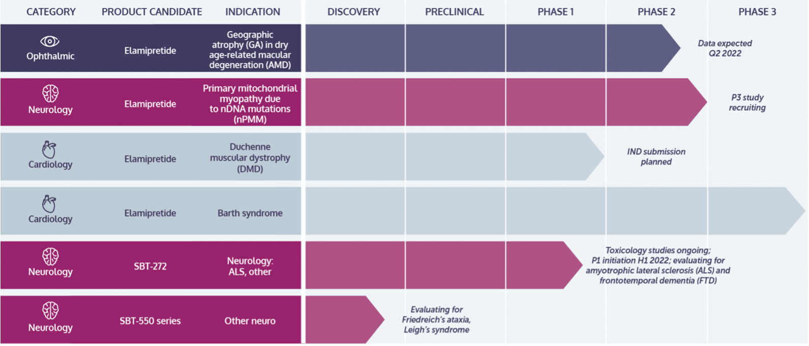
In November 2017 Stealth Peptides, Inc. of Newton, Mass, had obtained an orphan designation in the US for use in mitochondrial myopathy and had started a Phase III trial in that indication. As of January 2020, trial expectations were not met.
Elamipretide is currently also being tested in primary mitochondrial myopathy, for which it recently received an orphan-drug designation from the European Medicines Agency.
[i] Szeto HH. First-in-class cardiolipin-protective compound as a therapeutic agent to restore mitochondrial bioenergetics. Br J Pharmacol. 2014;171(8):2029-50.
[ii] Birk AV, et al. The mitochondrial-targeted compound SS-31 re-energizes ischemic mitochondria by interacting with cardiolipin. J Am Soc Nephrol. 2013;24(8):1250-61.
[iii] Szeto HH. First-in-class cardiolipin-protective compound as a therapeutic agent to restore mitochondrial bioenergetics. Br J Pharmacol. 2014;171(8):2029-50.
[iv] Wu J, et al. BDNF pathway is involved in the protective effects of SS-31 on isoflurane-induced cognitive deficits in aging mice. Behav Brain Res. 2016;305:115-21.
[v]Saad A, et al. Phase 2a Clinical Trial of Mitochondrial Protection (Elamipretide) During Stent Revascularization in Patients With Atherosclerotic Renal Artery Stenosis. Circ Cardiovasc Interv. 2017;10(9):e005487.
Clinical Research
IPS Level of Evidence
IPS Clinical Pharmacists have developed a method of ranking the studies so that the practitioner can easily discern the level of evidence this study provides to the topic. Levels 1-8 are listed below:
| Level of Evidence | Description | |
| X | Level 1 | FDA Approved Drug studies |
| Level 2 | Evidence obtained from systematic review and/or meta-analyses of studies including RCTs and other human studies | |
| X | Level 3 | Evidence obtained from a RCT |
| Level 4 | Evidence obtained from a study without randomization | |
| Level 5 | Evidence obtained from case reports | |
| X | Level 6 | Evidence obtained from in vitro human studies |
| X | Level 7 | Evidence obtained from laboratory animal studies |
| X | Level 8 | Evidence obtained from Opinions or Reviews |
Levels
Level 1
Saad A, et al. Phase 2a Clinical Trial of Mitochondrial Protection (Elamipretide) During Stent Revascularization in Patients With Atherosclerotic Renal Artery Stenosis. Circ Cardiovasc Interv. 2017;10(9):e005487.
Abstract
Background: Atherosclerotic renal artery stenosis reduces renal blood flow (RBF) and amplifies stenotic kidney hypoxia. Revascularization with percutaneous transluminal renal angioplasty (PTRA) and stenting often fails to recover renal function, possibly because of ischemia/reperfusion injury developing after PTRA. Elamipretide is a mitochondrial-targeted peptide that binds to cardiolipin and stabilizes mitochondrial function. We tested the hypothesis that elamipretide plus PTRA would improve renal function, oxygenation, and RBF in patients with atherosclerotic renal artery stenosis undergoing PTRA.
Methods and results: Inpatient studies were performed in patients with severe atherosclerotic renal artery stenosis scheduled for PTRA. Patients were treated before and during PTRA with elamipretide (0.05 mg/kg per hour intravenous infusion, n=6) or placebo (n=8). Stenotic kidney cortical/medullary perfusion and RBF were measured using contrast-enhanced multidetector CT, and renal oxygenation by 3-T blood oxygen level-dependent magnetic resonance imaging before and 3 months after PTRA. Age and basal glomerular filtration rate did not differ between groups. Blood oxygen level-dependent imaging demonstrated increased fractional hypoxia 24 hours after angiography and stenting in placebo (+47%) versus elamipretide (-6%). These were reverted to baseline 3 months later. Stenotic kidney RBF rose (202±29-262±115 mL/min; P=0.04) 3 months after PTRA in the elamipretide-treated group only. Over 3 months, systolic blood pressure decreased, and estimated glomerular filtration rate increased (P=0.003) more in the elamipretide group than in the placebo group (P=0.11).
Conclusions: Adjunctive elamipretide during PTRA was associated with attenuated postprocedural hypoxia, increased RBF, and improved kidney function in this pilot trial. These data support a role for targeted mitochondrial protection to minimize procedure-associated ischemic injury and to improve outcomes of revascularization for human atherosclerotic renal artery stenosis.
https://www.ncbi.nlm.nih.gov/pmc/articles/PMC5659347/pdf/nihms895904.pdf
Gibson CM, et al. EMBRACE STEMI study: a Phase 2a trial to evaluate the safety, tolerability, and efficacy of intravenous MTP-131 on reperfusion injury in patients undergoing primary percutaneous coronary intervention. Eur Heart J. 2016;37(16):1296-303.
Abstract
Aims: Among patients with ST-elevation myocardial infarction (STEMI), reperfusion injury contributes to additional myocardial damage. MTP-131 is a cell-permeable peptide that preserves the integrity of cardiolipin, enhances mitochondrial energetics, and improves myocyte survival during reperfusion.
Methods and results: EMBRACE STEMI is a multicentre, randomized, double-blind Phase 2a trial that evaluated the efficacy and safety of MTP-131 vs. placebo infused at a rate of 0.05 mg/kg/h for 1 h among first-time anterior STEMI subjects undergoing primary percutaneous coronary intervention (PCI) for a proximal or mid left anterior descending (LAD) artery occlusion. Administration of MTP-131 was not associated with a significant reduction in the primary endpoint, infarct size by creatine kinase-myocardial band (CK-MB) area under the curve (AUC) over 72 h (5785 ± 426 ng h/mL in placebo vs. 5570 ± 486 ng h/mL in MTP-131; ITALIC! P = NS). MTP-131 was not associated with an improvement in pre-specified magnetic resonance imaging, angiographic, electrocardiographic, or clinical outcomes.
Conclusion: Among subjects with first-time anterior STEMI due to a proximal or mid LAD lesion who undergo successful PCI, administration of MTP-131 was safe and well tolerated. Treatment with MTP-131 was not associated with a decrease in myocardial infarct size as assessed by AUC0-72 of CK-MB.
https://sci-hub.se/10.1093/eurheartj/ehv597
Thompson WR, et al. A phase 2/3 randomized clinical trial followed by an open-label extension to evaluate the effectiveness of elamipretide in Barth syndrome, a genetic disorder of mitochondrial cardiolipin metabolism. Genet Med. 2021;23(3):471-78.
Abstract
Purpose: To evaluate effectiveness of elamipretide in Barth syndrome (BTHS), a genetic condition of defects in TAZ, which causes abnormal cardiolipin on the inner mitochondrial membrane.
Methods: We performed a randomized, double-blind, placebocontrolled crossover trial followed by an open-label extension in BTHS to test the effect of elamipretide, a mitochondrial tetrapeptide that interacts with cardiolipin. In part 1, 12 subjects were randomized to 40 mg per day of elamipretide or placebo for 12 weeks, followed by a 4-week washout and then 12 weeks on the opposite arm. Ten subjects continued on the open-label extension (part 2) of 40 mg per day of elamipretide, with eight subjects reaching 36 weeks. Primary endpoints were improvement on the 6-minute walk test (6MWT) and improvement on a BTHS Symptom Assessment (BTHS-SA) scale.
Results: In part 1 neither primary endpoint was met. At 36 weeks in part 2, there were significant improvements in 6MWT (+95.9 m, p = 0.024) and BTHS-SA (-2.1 points, p = 0.031). There were also significant improvements in secondary endpoints including knee extensor strength, patient global impression of symptoms, and some cardiac parameters.
Conclusion: In this interventional clinical trial in BTHS, daily administration of elamipretide led to improvement in BTHS symptoms.
https://www.ncbi.nlm.nih.gov/pmc/articles/PMC7935714/pdf/41436_2020_Article_1006.pdf
Level 3
Karaa A, et al. A randomized crossover trial of elamipretide in adults with primary mitochondrial myopathy. J Cachexia Sarcopenia Muscle. 2020;11:909-18.
Abstract
Background This study aims to evaluate the effect of subcutaneous (SC) elamipretide dosing on exercise performance using the 6 min walk test (6MWT), patient-reported outcomes measuring fatigue, functional assessments, and safety to guide the development of the Phase 3 trial.
Methods MMPOWER-2 was a randomized, double-blind, placebo-controlled, crossover trial that enrolled participants (N = 30) with genetically confirmed primary mitochondrial myopathy. Participants were randomly assigned (1:1) to 40 mg/day SC elamipretide for 4 weeks followed by placebo SC for 4 weeks, separated by a 4-week washout period, or the opposite sequence. The primary endpoint was the distance walked on the 6MWT.
Results The distance walked on the 6MWT by the elamipretide-treated participants was 398.3 (±134.16) meters compared with 378.5 (±125.10) meters in the placebo-treated group, a difference of 19.8 m (95% confidence interval, 2.8, 42.5; P = 0.0833). The results of the Primary Mitochondrial Myopathy Symptom Assessment Total Fatigue and Total Fatigue During Activities scores showed that participants treated with elamipretide reported less fatigue and muscle complaints compared with placebo (P = 0.0006 and P = 0.0018, respectively). Additionally, the Neuro-QoL Fatigue Short Form and Patient Global Assessment showed reductions in symptoms (P = 0.0115 and P = 0.0421, respectively). In this 4-week treatment period, no statistically significant change was observed in the Physician Global Assessment (P = 0.0636), the Triple Timed Up and Go (P = 0.8423) test, and wrist/hip accelerometry (P = 0.9345 and P = 0.7326, respectively). Injection site reactions were the most commonly reported adverse events with elamipretide (80%), the majority of which were mild. No serious adverse events or deaths were reported.
Conclusions Participants who received a short-course treatment of daily SC elamipretide for 4 weeks experienced a clinically meaningful change in the 6MWT, which did not achieve statistical significance as the primary endpoint of the study. Secondary endpoints were suggestive of an elamipretide treatment effect compared with placebo. Nominal statistically significant and clinically meaningful improvements were seen in patient-reported outcomes. The results of this trial provided an efficacy signal and data to support the initiation of MMPOWER-3, a 6-month long, Phase 3 treatment trial in patients with primary mitochondrial myopathy.
https://www.ncbi.nlm.nih.gov/pmc/articles/PMC7432581/pdf/JCSM-11-909.pdf
Butler J, et al. Effects of Elamipretide on Left Ventricular Function in Patients With Heart Failure With Reduced Ejection Fraction: The PROGRESS-HF Phase 2 Trial. J Card Fail. 2020;26(5):429-37.
Abstract
Background: Elamipretide, a novel mitochondrial modulating agent, improves myocardial energetics; however, it is unknown whether this mechanistic benefit translates into improved cardiac structure and function in heart failure (HF) with reduced ejection fraction (HFrEF). The objective of this study was to evaluate the effects of multiple subcutaneous doses of elamipretide on left ventricular end systolic volume (LVESV) as assessed by cardiac magnetic resonance imaging.
Methods: We randomized 71 patients with HFrEF (LVEF ≤ 40%) in a double-blind, placebo-controlled trial in a 1:1:1 ratio to receive placebo, 4 mg or 40 mg elamipretide once daily for 28 consecutive days.
Results: The mean age (standard deviation) of the study population was 65 ± 10 years, 24% were females, and the mean EF was 31% ± 7%. The change in LVESV from baseline to week 4 was not significantly different between elamipretide 4 mg (89.4 mL to 85 mL; difference, -4.4 mL) or 40 mg (77.9 mL to 76.6 mL; difference, -1.2 mL) compared with placebo (77.7 mL to 74.6 mL; difference, -3.8 mL) (4 mg vs placebo: difference of means, -0.3; 95% CI, -4.6 to 4.0; P = 0.90; and 40 mg vs placebo: difference of means, 2.3; 95% CI, -1.9 to 6.5; P = 0.28). Also, no significant differences in change in LVESV and LVEF were observed between placebo and either of the elamipretide groups. Rates of any study drug-related adverse events were similar in the 3 groups.
Conclusions: Elamipretide was well tolerated but did not improve LVESV at 4 weeks in patients with stable HFrEF compared with placebo.
Level 6
Escribano-Lopez I, et al. The mitochondrial antioxidant SS-31 increases SIRT1 levels and ameliorates inflammation, oxidative stress and leukocyte-endothelium interactions in type 2 diabetes.
Abstract
There is growing focus on mitochondrial impairment and cardiovascular diseases (CVD) in type 2 diabetes (T2D), and the development of novel therapeutic strategies in this context. It is unknown whether mitochondrial-targeting antioxidants such as SS-31 protect sufficiently against oxidative damage in diabetes. We aimed to evaluate if SS-31 modulates SIRT1 levels and ameliorates leukocyte-endothelium interactions, oxidative stress and inflammation in T2D patients. Anthropometric and metabolic parameters were studied in 51 T2D patients and 57 controls. Production of mitochondrial reactive oxygen species (ROS), mitochondrial membrane potential, glutathione content, leukocyte-endothelium interactions, NFκB-p65, TNFα and SIRT1 levels was measured in leukocytes treated or not with SS-31. We observed increased mitochondrial ROS production that was restored by SS-31 treatment. SS-31 also increased mitochondrial membrane potential, glutathione content, SIRT1 levels and leukocyte rolling velocity and reduced rolling flux and adhesion in T2D patients. NFκB-p65 and TNFα, which were enhanced in diabetic patients, were also reduced by SS-31 treatment. Our results reveal that SS-31 exerts beneficial effects on the leukocytes of T2D patients by reducing oxidative stress, leukocyte-endothelium interactions, NFκB and TNFα and by increasing SIRT1 levels. These actions support its use as a potential agent against CVD risk.
https://www.nature.com/articles/s41598-018-34251-8
Chen M, et al. Protective effect of mitochondria-targeted peptide MTP-131 against oxidative stress-induced apoptosis in RGC-5 cells. Mol Med Rep. 2017;15(4):2179-85.
Abstract
The retina of the human eye is extremely vulnerable to oxidative damage. Previous studies have demonstrated that oxidative stress is the predominant mechanism associated with the pathogenesis of age-related macular degeneration, diabetic retinopathy, glaucoma and retinitis pigmentosa. MTP-131, a novel mitochondria-targeted peptide, has been demonstrated to specifically concentrate in the inner mitochondria membrane and to exhibit remarkable antioxidant effects both in vitro and in animal models. In the present study, the protective effect of MTP-131 was evaluated in response to hydrogen peroxide (H2O2)-induced oxidative damage in a retinal ganglion cell line, RGC-5. Cell viability was measured by lactate dehydrogenase (LDH) assay. Changes of mitochondrial membrane potential and generation of intracellular reactive oxygen species (ROS) were measured by flow cytometry and confocal microscopy, respectively. Annexin V-fluorescein isothiocyanate/propidium iodide staining was used for assessment of apoptosis. Release of cytochrome c was analyzed by confocal microscopy. Pretreatment of cells with MTP-131 inhibited H2O2-induced cytotoxicity and reduced LDH release in a dose-dependent manner, compared with cells treated with H2O2 alone. Mitochondrial depolarization and ROS generation were also prevented by MTP-131 pretreatment. In addition, MTP-131 pretreatment inhibited cytochrome c release from mitochondria to cytoplasm, and significantly reduced apoptosis in RGC-5 cells, compared with cells treated with H2O2 alone. In conclusion, mitochondria-targeted peptide MTP-131 exhibited a protective effect against oxidative stress-induced apoptosis in RGC-5 cells, which may provide a novel approach for the treatment of age-associated retinal diseases.
https://www.ncbi.nlm.nih.gov/pmc/articles/PMC5364865/pdf/mmr-15-04-2179.pdf
Level 7
Wu X, et al. Mitochondria-targeted antioxidant peptide SS-31 mediates neuroprotection in a rat experimental glaucoma model. Acta Biochim Biophys Sin (Shanghai). 2019;51(4):411-21.
Abstract
To investigate the neuroprotective effects of the mitochondria-targeted antioxidant Szeto-Schiller peptide 31 (SS-31) in a rat experimental glaucoma model, SS-31 was intraperitoneally (IP) injected into Sprague-Dawley rats, followed by intracameral injection of polystyrene microspheres to induce elevated intraocular pressure (IOP). After 6 weeks, electroretinography (ERG) and flash visual-evoked potentials (F-VEPs) were recorded to assess retinal function. Hematoxylin-eosin staining was performed on retinal cross-sections to measure ganglion cell complex (GCC) thickness. Apoptotic retinal cells were assessed by TUNEL staining. Brn3a-positive retinal ganglion cells (RGCs) were counted in retinal flat mounts via immunofluorescence. The retinal total SOD, SOD2, and MDA expression levels were assessed in retinal tissue homogenates. The cyt c, Bax, and Bcl-2 protein levels in rat retinas were detected by western blot analysis. Bax and Bcl-2 expressions were also evaluated using immunohistochemistry in paraffinized sections. Our results showed that the rats that received microsphere injection developed elevated IOP. SS-31 ameliorated the reductions in the a- and b-wave amplitudes on ERG and the F-VEP amplitude in glaucomatous eyes. GCC thickness was preserved, TUNEL-positive cells were decreased in the retina, and Brn3a-positive RGCs were increased in the SS-31-treated glaucoma group compared with those in the non-treated glaucoma group. SS-31 significantly reduced MDA levels and increased SOD2 levels after glaucoma induction. Significant suppression of cyt c release, upregulation of Bcl-2, and downregulation of Bax were observed following SS-31 administration. In summary, SS-31 exerts neuroprotective effects in this experimental glaucoma model by inhibiting mitochondrial dysfunction and therefore represents a promising therapeutic agent for glaucoma.
https://sci-hub.se/10.1093/abbs/gmz020
Zhao W, et al. Elamipretide (SS-31) improves mitochondrial dysfunction, synaptic and memory impairment induced by lipopolysaccharide in mice. J Neuroinflam. 2019;16:230.
Abstract
Background: It is widely accepted that mitochondria have a direct impact on neuronal function and survival. Oxidative stress caused by mitochondrial abnormalities play an important role in the pathophysiology of lipopolysaccharide (LPS)-induced memory impairment. Elamipretide (SS-31) is a novel mitochondrion-targeted antioxidant. However, the impact of elamipretide on the cognitive sequelae of inflammatory and oxidative stress is unknown.
Methods: We utilized MWM and contextual fear conditioning test to assess hippocampus-related learning and memory performance. Molecular biology techniques and ELISA were used to examine mitochondrial function, oxidative stress, and the inflammatory response. TUNEL and Golgi-staining was used to detect neural cell apoptosis and the density of dendritic spines in the mouse hippocampus.
Results: Mice treated with LPS exhibited mitochondrial dysfunction, oxidative stress, an inflammatory response, neural cell apoptosis, and loss of dendritic spines in the hippocampus, leading to impaired hippocampus-related learning and memory performance in the MWM and contextual fear conditioning test. Treatment with elamipretide significantly ameliorated LPS-induced learning and memory impairment during behavioral tests. Notably, elamipretide not only provided protective effects against mitochondrial dysfunction and oxidative stress but also facilitated the regulation of brain-derived neurotrophic factor (BDNF) signaling, including the reversal of important synaptic-signaling proteins and increased synaptic structural complexity.
Conclusion: These findings indicate that LPS-induced memory impairment can be attenuated by the mitochondrion-targeted antioxidant elamipretide. Consequently, elamipretide may have a therapeutic potential in preventing damage from the oxidative stress and neuroinflammation that contribute to perioperative neurocognitive disorders (PND), which makes mitochondria a potential target for treatment strategies for PND.
https://sci-hub.se/10.1186/s12974-019-1627-9
Zuo Y, et al. Elamipretide Attenuates Pyroptosis and Perioperative Neurocognitive Disorders in Aged Mice. Front Cell Neurosci. 2020;14:251.
Abstract
Pyroptosis is a recently characterized inflammatory form of programmed cell death that is thought to be involved in the pathogenesis of perioperative neurocognitive disorders (PND). Elamipretide (SS-31), a mitochondrial-targeted peptide with multiple pharmacological properties, including anti-inflammatory activity, has been demonstrated to protect against many neurological diseases. However, the effect of elamipretide on pyroptosis in PND has not been studied. We established an animal model of PND by performing an exploratory laparotomy on mice under isoflurane anesthesia and examined the effects of elamipretide on cognitive function, synaptic integrity, neuroinflammation, mitochondrial function, and signaling controlling pyroptosis. Our results showed that anesthesia and surgery caused mitochondrial dysfunction and abnormal morphology, activation of canonicalnod-like receptor pyrin domain-containing 3 (NLRP3) inflammasome-caspase-1 dependent pyroptosis, and downregulation of synaptic integrity-related proteins in the hippocampus in aged mice, thus leading to learning and memory deficits in behavioral tests. Remarkably, treatment with the mitochondrial-targeted peptide elamipretide not only had protective effects against mitochondrial dysfunction but also attenuated surgery-induced pyroptosis and cognitive deficits. Our results provide a promising strategy for the treatment of PND involving mitochondrial dysfunction and pyroptosis.
https://www.ncbi.nlm.nih.gov/pmc/articles/PMC7439217/pdf/fncel-14-00251.pdf
Dai W, et al. Bendavia, a mitochondria-targeting peptide, improves postinfarction cardiac function, prevents adverse left ventricular remodeling, and restores mitochondria-related gene expression in rats. J Cardiovasc Pharmacol. 2014;64(6):543-53.
Abstract
AB We evaluated the post-myocardial infarction (MI) therapeutic effects of Bendavia. Two hours after coronary artery ligation, rats were randomized to receive chronic Bendavia treatment (n = 28) or water (n = 26). Six weeks later, Bendavia significantly reduced scar circumference (39.7% +/- 2.2%) compared with water treatment (47.4% +/- 0.03%, P = 0.024) and reduced left ventricular (LV) volume by 8.9% (P = 0.019). LV fractional shortening was significantly improved by Bendavia (28.8% +/- 1.7%) compared with water treatment (23.8% +/- 1.8%, P = 0.047). LV ejection fraction was higher with Bendavia (55.3% +/- 1.4%) than water treatment (49.3% +/- 1.4%, P = 0.005). Apoptosis, within the MI border zone, was significantly less in the Bendavia group (32% +/- 3%, n = 12) compared with the water group (41% +/- 2%, n = 12; P = 0.029). Bendavia reversed mitochondrial function-related gene expression in the MI border, which was largely reduced in water-treated rats. Bendavia improved complex-I and -IV activity, and reduced production of reactive oxygen species and cytosolic cytochrome c level in the peri-infarcted region. Bendavia improved post-MI cardiac function, prevented infarct expansion and adverse LV remodeling, and restored mitochondria-related gene expression, complex-I and -IV activity, and reduced reactive oxygen species and cardiomyocyte apoptosis in the noninfarcted MI border.
https://sci-hub.se/10.1097/fjc.0000000000000155
Bartell LR, et al. The cardiolipin-stabilizing peptide SS-31 protects chondrocytes from peracute, mechanically-dependent mitochondrial depolarization and preserves christae structure after cartilage injury. Osteoarth Cartilage. 2018;26(1):S63-S64.
Abstract
Purpose: Understanding and targeting cellular events that occur immediately after cartilage injury may lead to effective disease-modifying osteoarthritis (OA) therapies that preserve cartilage homeostasis and prevent perpetual cell death and matrix degradation. Increasing evidence implicates mitochondrial (MT) dysfunction in the initiation and early pathogenesis of post-traumatic OA. Acute MT respiratory dysfunction has been identified within hours after cartilage impact-injury, and targeted mitoprotection administered up to 6 hours after injury prevented chondrocyte death, apoptosis and matrix degradation. Szeto-Schiller (SS) peptides prevent MT dysfunction by stabilizing cardiolipin, a phospholipid essential for formation of cristae structure and electron transport chain super-complexes on the inner MT membrane. The goals of this study were to (1) investigate the mechanical dependence of MT depolarization immediately after cartilage impact (i.e., peracute time frame), and (2) test if a mitoprotective peptide (SS-31) can preserve MT structure and function.
Methods: Cartilage explants were harvested from femoral condyles of neonatal calves, bisected, and mounted to a mechanical test frame (n = 7 animals; Fig. 1A). One hemicylinder was impacted rapidly (∼1 MPa over 5–10 ms) while the other served as an uninjured control. Some samples (n = 3) were treated with SS-31 (1 uM) prior to injury. All explants were stained with fluorescent probes to identify three cell populations: live cells with polarized/functional MT, live cells with depolarized MT, and dead cells. Explants were imaged longitudinally using confocal microscopy before and up to 60 minutes after impact, in various locations relative to the impact (Fig. 1B). Images were processed using MATLAB to segment individual cells and track their fate over time (Fig. 1C). Local sample deformation during impact was determined using an FEBio model, which was validated by fast-camera images of actual sample deformation. Electron microscopy was used to evaluate MT structure at 30 minutes after impact.
Results: A rapid wave of chondrocyte MT depolarization occured within 3 minutes after impact, then progressed more slowly over course of the experiment. SS-31 treatment reduced this peracute depolarization (Fig. 2AB). Without treatment, the average fraction of cells with depolarized MT was highest near the impact, decreased with distance from the impact, and was highly correlated with local tissue strain (Fig. 2CD). In contrast, treatment with SS-31 prevented this strain-dependent MT depolarization (Fig. 2EF). Without treatment, electron microscopy revealed impact-induced MT swelling and loss of cristae, while SS-31 treatment preserved cristae structure after impact.
https://sci-hub.se/10.1016/j.joca.2018.02.135
Mo Y, et al. SS-31 reduces inflammation and oxidative stress through the inhibition of Fis1 expression in lipopolysaccharide-stimulated microglia. Biochem Biophys Res Commun. 2019;520(1):171-78.
Abstract
SS-31 is a kind of mitochondrion-targeted peptide. Recent studies indicated significant neuroprotective effects of SS-31. In this study, we investigated that SS-31 protected the murine cultured microglial cells (BV-2) against lipopolysaccharide (LPS)-induced inflammation and oxidative stress through stabilizing mitochondrial morphology. The morphological study showed that SS-31 preserved LPS-induced mitochondrial ultrastructure by reducing the fission protein 1 (Fis1) expression. Flow cytometry and Western blot verified that SS-31 defended the BV-2 cells against LPS-stimulated inflammation and oxidative stress via suppressing Fis1. To sum up, our study represents that SS-31 preserves BV-2 cells from LPS-stimulated inflammation and oxidative stress by down-regulating the Fis1 expression.
https://sci-hub.se/10.1016/j.bbrc.2019.09.077
Birk AV, et al. The mitochondrial-targeted compound SS-31 re-energizes ischemic mitochondria by interacting with cardiolipin. J Am Soc Nephrol. 2013;24(8):1250-61.
Abstract
Ischemia causes AKI as a result of ATP depletion, and rapid recovery of ATP on reperfusion is important to minimize tissue damage. ATP recovery is often delayed, however, because ischemia destroys the mitochondrial cristae membranes required for mitochondrial ATP synthesis. The mitochondria-targeted compound SS-31 accelerates ATP recovery after ischemia and reduces AKI, but its mechanism of action remains unclear. Here, we used a polarity-sensitive fluorescent analog of SS-31 to demonstrate that SS-31 binds with high affinity to cardiolipin, an anionic phospholipid expressed on the inner mitochondrial membrane that is required for cristae formation. In addition, the SS-31/cardiolipin complex inhibited cytochrome c peroxidase activity, which catalyzes cardiolipin peroxidation and results in mitochondrial damage during ischemia, by protecting its heme iron. Pretreatment of rats with SS-31 protected cristae membranes during renal ischemia and prevented mitochondrial swelling. Prompt recovery of ATP on reperfusion led to rapid repair of ATP-dependent processes, such as restoration of the actin cytoskeleton and cell polarity. Rapid recovery of ATP also inhibited apoptosis, protected tubular barrier function, and mitigated renal dysfunction. In conclusion, SS-31, which is currently in clinical trials for ischemia-reperfusion injury, protects mitochondrial cristae by interacting with cardiolipin on the inner mitochondrial membrane.
https://www.ncbi.nlm.nih.gov/pmc/articles/PMC3736700/
Wu J, et al. BDNF pathway is involved in the protective effects of SS-31 on isoflurane-induced cognitive deficits in aging mice. Behav Brain Res. 2016;305:115-21.
Abstract
Mitochondrial dysfunction has been linked to the earliest pathogenesis of isoflurane-induced cognitive impairments in developing or aging mammalian brain. However, its molecular mechanism is poorly understood and a pharmacologic treatment to rapidly reverse mitochondrial dysfunction is lacking. Fifteen-month-old male C57BL/6 mice were exposed to isoflurane for two hours following intraperitoneal administration of mitochondrion-targeted peptide SS-31 or vehicle with 30min interval. The hippocampus was immediately removed for biochemical assays and mitochondria isolation after inhalation. Behavioral tests were evaluated by the open field test and fear conditioning test 24h after the experiment. We showed that cognitive deficits induced by exposure of the aging mice to isoflurane were accompanied by mitochondrial dysfunction in hippocampus due to loss of the enzymatic activity of complex I. This loss resulted in the increase of reactive oxygen species production, decrease of ATP production and mitochondrial membrane potential, and opening of mitochondrial permeability transition pore. Further, we provided evidence that the BDNF signaling pathway was involved in this process to regulate synaptic plasticity-related proteins, for instance, downregulation of synapsin 1, PSD-95 and p-CREB, and upregulation of NR2A, NR2B, CaMKIIα and CaMKIIβ. Of note, the isoflurane-induced cognitive deficits were rescued by SS-31 through reversal of mitochondrial dysfunction, which facilitated the regulation of BDNF signaling including the expression reversal of aforementioned important synaptic-signaling proteins in aging mice. Our data demonstrate that reversing mitochondrial dysfunction by SS-31 enhances BDNF signaling pathway and synaptic plasticity, and provides protective effects on cognitive function, thereby support the notion that SS-31 may have therapeutic benefits for elderly humans undertaking anesthesia.
Level 8
Szeto HH. First-in-class cardiolipin-protective compound as a therapeutic agent to restore mitochondrial bioenergetics. Br J Pharmacol. 2014;171(8):2029-50.
Abstract
decline in energy is common in aging, and the restoration of mitochondrial bioenergetics may offer a common approach for the treatment of numerous age-associated diseases. Cardiolipin is a unique phospholipid that is exclusively expressed on the inner mitochondrial membrane where it plays an important structural role in cristae formation and the organization of the respiratory complexes into supercomplexes for optimal oxidative phosphorylation. The interaction between cardiolipin and cytochrome c determines whether cytochrome c acts as an electron carrier or peroxidase. Cardiolipin peroxidation and depletion have been reported in a variety of pathological conditions associated with energy deficiency, and cardiolipin has been identified as a target for drug development. This review focuses on the discovery and development of the first cardiolipin-protective compound as a therapeutic agent. SS-31 is a member of the Szeto-Schiller (SS) peptides known to selectively target the inner mitochondrial membrane. SS-31 binds selectively to cardiolipin via electrostatic and hydrophobic interactions. By interacting with cardiolipin, SS-31 prevents cardiolipin from converting cytochrome c into a peroxidase while protecting its electron carrying function. As a result, SS-31 protects the structure of mitochondrial cristae and promotes oxidative phosphorylation. SS-31 represents a new class of compounds that can recharge the cellular powerhouse and restore bioenergetics. Extensive animal studies have shown that targeting such a fundamental mechanism can benefit highly complex diseases that share a common pathogenesis of bioenergetics failure. This review summarizes the mechanisms of action and therapeutic potential of SS-31 and provides an update of its clinical development programme.
https://www.ncbi.nlm.nih.gov/pmc/articles/PMC3976620/pdf/bph0171-2029.pdf